
At a booking desk, during a prenatal visit, or on a clinic flyer, you may spot “3D/4D ultrasound available” and wonder what the numbers mean and whether the extra dimensions matter for your baby.
The short version: the difference comes down to how sound waves are assembled into a picture, not different machines firing different rays.
This guide explains what each type shows, when each scan is usually used, how 3D/4D ultrasound can fit into a personalized pregnancy care experience, and how to decide which option you actually need.
Quick Answer: The Difference Between 2D, 3D, and 4D Ultrasound During Pregnancy
Think of it as one technology with three different ways of showing the image. A 2D ultrasound is the standard flat, black-and-white scan used to check fetal growth, anatomy, heartbeat, placenta, fluid, and pregnancy dating.
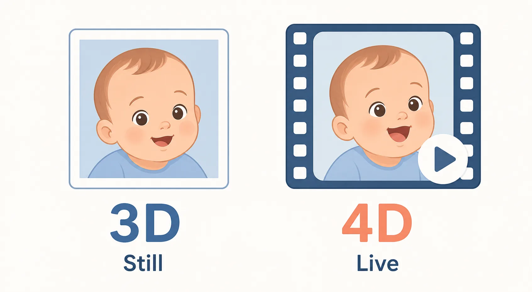
A 3D ultrasound creates a still three-dimensional image, often showing the baby’s face or surface features in more detail. A 4D ultrasound shows that 3D image in motion, so parents may see the baby yawn, stretch, smile, or move a hand near the face.
All three rely on high-frequency sound waves. What changes is how the software turns the returning echoes into an image.
At Minnesota Women’s Care, 4D ultrasound is available as part of a broader prenatal care experience. Obstetric patients may receive a 4D image during the routine anatomy scan, and additional 4D ultrasound session options are available for families who want more keepsake images or video-style memories.
| Scan type | What it shows | Best use | Usually needed? |
|---|---|---|---|
| 2D ultrasound | Flat black-and-white cross-sections | Dating pregnancy, anatomy, growth, organs, placenta, and fluid | Yes, for routine medical care |
| 3D ultrasound | Still three-dimensional surface image | Face, profile, fingers, and surface features | Sometimes; often optional |
| 4D ultrasound | 3D image in real-time motion | Yawns, stretches, facial movement, and bonding | Usually optional unless your provider recommends it |
| 5D/HD ultrasound | Enhanced rendering of 3D/4D data | More lifelike keepsake-style images | Not medically separate |
If your goal is medical information, 2D ultrasound is usually the scan you need. If your goal is bonding, keepsake images, or seeing your baby’s movements more clearly, 3D or 4D ultrasound may be worth asking about during prenatal care.
How 2D, 3D, and 4D Ultrasound Work

Ultrasound uses high-frequency sound waves to create images from inside the body. During pregnancy, those sound waves bounce back from fetal tissue, fluid, and surrounding structures. The ultrasound system then converts those returning echoes into images on a screen.
The basic technology is similar across 2D, 3D, and 4D ultrasound. What changes is the image processing, not the use of different rays or radiation.
2D ultrasound
A 2D ultrasound sends sound waves through the body and displays the returning echoes as flat, black-and-white cross-sections. These images may not look like a portrait, but they give clinicians the views they need to measure and assess the pregnancy.
That is why 2D ultrasound remains the standard for many medical questions during pregnancy, including fetal growth, anatomy, heartbeat, placenta location, and amniotic fluid.
3D ultrasound
A 3D ultrasound captures multiple 2D images from different angles and reconstructs them into a still three-dimensional image. Instead of seeing only a flat slice, parents may be able to see the curve of the baby’s cheek, nose, lips, or hands.
This is the type of ultrasound many families think of when they want a more lifelike keepsake image.
4D ultrasound
A 4D ultrasound takes rapid 3D images and displays them in real time. That means parents may see movement as it happens, such as a stretch, yawn, turn, or hand moving near the face. In simple terms, 4D ultrasound is 3D ultrasound with motion.
A review of three- and four-dimensional ultrasound in obstetric practice explains that 3D/4D imaging uses volume datasets that can be reviewed, rotated, and displayed from different perspectives.
That added processing can help visualize fetal surface features and movement, although 2D ultrasound remains the standard tool for many routine diagnostic measurements.
4D ultrasound is an advanced imaging technique that allows real-time visualization of the baby’s movements, facial expressions, and some …Read on LinkedIn ↗
What Each Pregnancy Ultrasound Scan Shows
Each ultrasound type has a different strength. One is not automatically “better” than the others. The right scan depends on whether your provider needs medical measurements, whether you want a more recognizable keepsake image, or whether both goals matter during your pregnancy care.
| Scan type | What it shows best | Main purpose |
|---|---|---|
| 2D ultrasound | Internal anatomy, growth, heartbeat, placenta, fluid, and pregnancy dating | Medical assessment |
| 3D ultrasound | Surface features such as the face, profile, hands, feet, and fingers | More lifelike still image |
| 4D ultrasound | Real-time movement, such as yawning, stretching, turning, or moving a hand | Motion view and bonding experience |
Diagnostic detail
2D ultrasound remains the clinical standard for most pregnancy imaging. It shows flat cross-sections that help your provider check fetal anatomy, growth, heartbeat, placenta location, amniotic fluid, and pregnancy dating.
These images may not look like a portrait of your baby, but that is not their purpose. For measuring anatomy, checking organs, and answering medical questions, flat cross-sectional views are often exactly what your provider needs.
Surface and facial features
3D ultrasound is often easier for parents to recognize because it shows the outside surface of the baby’s body. When the baby is in a good position, a 3D scan may show the face, profile, nose, lips, hands, feet, or fingers more clearly than a standard 2D image.
This is what many parents mean when they say they want to “see the baby.” It gives a still, more lifelike view, but it does not replace the medical value of a standard 2D scan.
Movement and bonding
4D ultrasound adds real-time motion to the 3D image. Instead of seeing one still picture, you may see the baby yawn, stretch, turn, smile, or move a hand near the face.
Many parents find this emotionally meaningful because it can make the pregnancy feel more personal and real. That bonding value matters, even when the scan does not change the medical plan.
That bonding value matters, even when the scan does not change the medical plan.
So, do 2D ultrasounds look like the baby? They show your baby, but not in a photo-like way. A 2D scan is designed to show cross-sections that help your provider evaluate the pregnancy, so that the image may look less recognizable to parents than a 3D or 4D view.
And are 3D and 4D clearer than 2D? They are usually clearer for surface details, such as the baby’s face or hands. For medical measurements, anatomy checks, and many diagnostic questions, 2D ultrasound remains the more useful tool.
At MWC Pregnancy, the goal is to keep that experience connected to provider-guided prenatal care, so parents can enjoy the keepsake side while still receiving thoughtful medical support.
When each type is used during pregnancy
2D is used throughout pregnancy, from early dating through the mid-pregnancy anatomy scan. 3D and 4D are usually elective add-ons, most often performed in the late second or early third trimester, when there is sufficient fetal development and fluid to capture facial detail.
Minnesota Women’s Care notes that the clearest images of the baby’s features are normally captured between 24 and 32 weeks, depending on the baby’s position and the amount of amniotic fluid around the baby.
Even in that ideal window, image quality can vary if the baby is turned away, covering the face, or positioned close to the placenta or uterine wall.
Occasionally a clinician uses 3D or 4D to assess a specific concern, such as a suspected facial or skeletal difference.
Can you ask your OB for a 4D ultrasound? You can raise it, but keep expectations realistic. OBs order scans for medical need; purely elective 3D and 4D imaging is often done at dedicated imaging centers rather than in the clinic.
Are 3D and 4D ultrasounds safe?
3D and 4D ultrasound use the same sound-wave technology used in standard pregnancy ultrasound. Ultrasound does not use ionizing radiation, which is one reason it is widely used in prenatal care.
The important point is how ultrasound is used. The FDA notes that ultrasound is not based on ionizing radiation, but it also discourages ultrasound used only for non-medical keepsake videos when it creates unnecessary or prolonged exposure. Keepsake images are considered more reasonable when captured during a medically indicated exam without adding extra exposure.
That is the balanced answer: ultrasound is commonly used in pregnancy care, but extra scans should still be thoughtful, purposeful, and performed by trained professionals.
How MWC Pregnancy Uses 3D/4D Ultrasound
Minnesota Women’s Care offers 3D/4D ultrasound as part of its pregnancy care experience. For obstetric patients, a 4D image is included with the routine anatomy scan, and additional 4D ultrasound packages are available for patients who want a longer session or more keepsake images.
This keeps the experience connected to prenatal care instead of treating ultrasound as a standalone novelty. The broader pregnancy care team includes nurse practitioners, nurse-midwives, and physicians, so patients can enjoy the bonding experience of 3D/4D imaging while staying connected to medical guidance.
For families comparing pregnancy care options, this is an important difference. A keepsake image can be special, but it feels more reassuring when it is part of a larger care plan that also includes prenatal visits, anatomy checks, labor and delivery support, postpartum care, and patient education.
Is a 4D ultrasound worth it?
For a low-risk pregnancy, a 4D scan usually adds keepsake and bonding value rather than medical value. If your care is going well, the deciding factor is how much that live glimpse means to you.
Cost tends to follow that split. A medically indicated 2D scan is typically billed as part of prenatal care, while elective 3D and 4D scans are usually paid out of pocket.
Prices vary widely by location and package, so confirm figures directly with the provider rather than assuming. If you are searching for a 2D, 3D, or 4D ultrasound near you, note that clinical scans happen in medical settings while elective ones run at imaging studios; either way, look for a trained sonographer and medical oversight.
What About 5D or HD Ultrasound?
You may also see terms like 5D, HD, or HD Live ultrasound. These usually refer to enhanced rendering, lighting, color, and shadow effects applied to 3D or 4D ultrasound images.
They can make the image look smoother or more lifelike, but they are not a completely new medical dimension. In most cases, 5D or HD ultrasound does not mean the scan gives your provider more medical information. It usually means the final image has a more polished visual appearance.
| Term | What it means | What parents usually notice |
|---|---|---|
| 3D ultrasound | A still three-dimensional surface image | A clearer view of the baby’s face or features |
| 4D ultrasound | A moving 3D image shown in real time | Yawns, stretches, hand movement, or facial movement |
| 5D/HD ultrasound | Enhanced rendering applied to 3D/4D data | Smoother lighting, shadows, and more lifelike skin tones |
So if you are comparing 3D, 4D, and 5D ultrasound, focus less on the label and more on who is performing the scan, whether there is medical oversight, how long the session lasts, and what you will actually receive.
How to Get the Clearest 3D/4D Ultrasound Images
A clear 3D or 4D image depends on several things, and not all of them are under your control.
The baby’s position matters most. If the baby is facing your back, covering the face, or pressed against the uterine wall, the image may not be clear. Amniotic fluid also matters because fluid around the face helps create a better view. Timing matters too, which is why many providers recommend the late second or early third trimester for the clearest facial images.
You can ask your provider what timing makes the most sense for your pregnancy, especially if you are hoping for a keepsake image.
Choosing the Right Ultrasound for Your Pregnancy
If you are trying to decide between 2D, 3D, and 4D ultrasound, start with your purpose.
If your goal is medical information, 2D ultrasound is usually the scan you need. If your goal is to see your baby’s face more clearly, 3D may offer that still-image view. If your goal is to see movement and create a more emotional keepsake, 4D may be the better fit.
For patients who want both reassurance and a more personal pregnancy experience, MWC Pregnancy’s approach brings these elements together: clinical prenatal care, on-site imaging, provider guidance, and the option of a more memorable 3D/4D ultrasound.
FAQs About Ultrasounds
Frequently asked questions
Are 3D and 4D ultrasounds clearer than 2D?
What’s the difference between 3D and 4D ultrasound?
What’s the difference between 3D, 4D, and 5D ultrasound?
Which is better, 3D or 4D ultrasound?
How much does a 3D/4D ultrasound cost?
Can you see baby hair in a 4D ultrasound?
Can too many ultrasounds harm a fetus?
Which is better, 2D or 4D?
Final Takeaway
The practical difference is simple: 2D ultrasound does most of the medical work, 3D ultrasound gives a still lifelike view, and 4D ultrasound adds motion.
If you are receiving pregnancy care through Minnesota Women’s Care, 4D imaging can be part of a broader prenatal experience rather than a separate souvenir scan. Talk with your care team about what you need medically, what you want emotionally, and which ultrasound option fits your pregnancy best.