
Halfway through pregnancy, you lie back for an ultrasound that takes a careful, head-to-toe look at your baby. This is the 20-week anatomy scan, a routine part of prenatal care. Most parents walk in wanting two things: to understand what the sonographer is actually checking, and to know what the results mean. Here is a plain-language guide to both.
Quick Answer: What Does a 20-Week Anatomy Scan Check?
A 20-week anatomy scan checks your baby’s brain, spine, face, heart, lungs, stomach, kidneys, bladder, abdominal wall, arms, legs, hands, feet, growth measurements, placenta position, umbilical cord, and amniotic fluid. It is a screening ultrasound, which means it can look for signs of certain structural concerns, but it cannot detect every possible condition.
What is a 20-week anatomy scan?
The 20-week anatomy scan is a detailed mid-pregnancy ultrasound, also called the anatomy ultrasound, fetal anatomy scan, or mid-pregnancy ultrasound. It is usually done between 18 and 22 weeks. The sonographer checks your baby’s growth, the development of the organs and body parts, the position of the placenta, and the amount of amniotic fluid.
ACOG says this second-trimester ultrasound is used to look for fetal structural defects and is ideally performed between 18 and 22 weeks of pregnancy. It can also help your care team decide whether you need a repeat view, follow-up ultrasound, genetic counseling, or another type of prenatal testing.
It helps to think of it as a screening tool rather than a guarantee. U.S. medical guidance describes ultrasound as a screening exam that can look for certain structural concerns, but it cannot detect every possible condition.
A reassuring scan is good news, but it is not a promise that nothing will ever be found later in pregnancy. Setting that expectation early makes the results easier to understand later.
When Is the Best Time for a 20-Week Anatomy Scan?
The anatomy scan is commonly scheduled around 18 to 22 weeks of pregnancy, depending on your provider’s recommendation and appointment availability. This timing gives the sonographer enough detail to see many of the baby’s organs and structures, while the baby is usually still small enough to view clearly in one exam.
If the scan is delayed or not completed the first time, your provider may recommend another detailed ultrasound or follow-up imaging. The right timing depends on your pregnancy, your clinic’s protocol, and what still needs to be checked.
A repeat scan does not automatically mean something is wrong. Sometimes the baby is curled up, facing the wrong direction, moving a lot, or blocking a view of the heart, spine, face, or other structures.
What Happens During the 20-Week Anatomy Scan?
During the scan, you usually lie on an exam table while gel is placed on your abdomen. The sonographer moves an ultrasound probe across your belly to capture different views of your baby. You may feel light pressure as they adjust the angle, especially when they need a clearer view of a specific area.
The appointment often takes around 30 minutes, but it can take longer if the baby’s position makes certain views harder to capture. You may be asked to change position, roll slightly, take a short break, or come back another day if the checklist cannot be completed.
The sonographer may point out some visible features, but they may not be able to explain every measurement or finding in detail during the exam. Your provider reviews the results and explains what was seen, whether the scan was complete, and whether any follow-up is needed.
What Does a 20-Week Anatomy Scan Check?
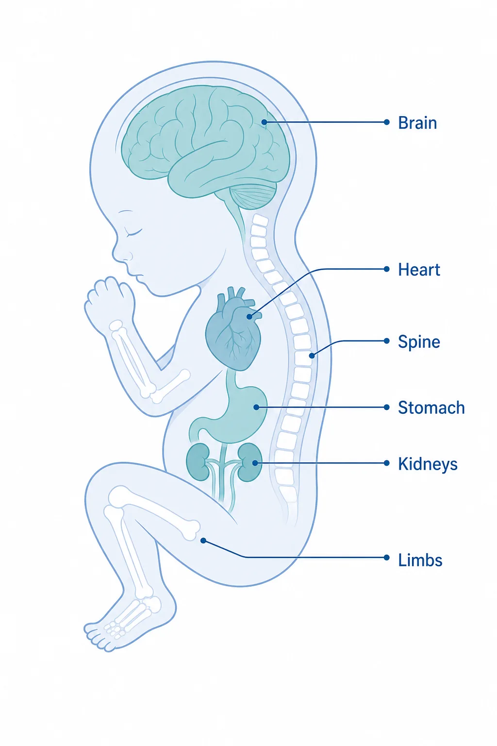
The sonographer works methodically through the body, and this is where the most detail lives. A second-trimester ultrasound guide published in the National Library of Medicine walks through the same structures organ by organ.
| Area checked | What the sonographer looks for |
|---|---|
| Head and brain | Skull shape, brain structures, and signs that development appears on track |
| Face | Profile, lips, and visible facial structures, including a look for signs of cleft lip |
| Spine | Vertebrae and the skin covering the spine, which helps screen for spinal concerns |
| Heart | Four chambers, rhythm, position, and major views of blood flow |
| Chest and lungs | Chest structure and lung development |
| Abdomen | Stomach, kidneys, bladder, bowel, and abdominal wall |
| Arms, legs, hands, and feet | Long bones, limb movement, and visible hands or feet when the baby’s position allows |
| Placenta and cord | Placenta position, cord insertion, and blood flow where assessed |
| Amniotic fluid | Whether the fluid level appears within the expected range |
| Growth measurements | Head circumference, abdominal circumference, and femur length |
The heart is often one of the most detailed parts of the exam. If the sonographer spends extra time trying to capture heart views, it does not automatically mean there is a problem. The baby may simply be moving, turned away, or in a position that makes the heart harder to see clearly.
The scan may also check the uterus, cervix, ovaries, or other maternal structures when clinically appropriate. If the placenta appears low, your provider may recommend another ultrasound later in pregnancy to see whether it has moved upward as the uterus grows.
Can a 20-Week Anatomy Scan Detect Down Syndrome?
A 20-week anatomy scan is not a diagnostic test for Down syndrome. It can sometimes show “soft markers,” which are small ultrasound findings that may slightly raise or lower the chance of a chromosome condition.
Soft markers can also appear in healthy pregnancies, so they are usually interpreted alongside your age, medical history, screening results, and any other ultrasound findings.
Screening tests estimate risk; diagnostic tests can confirm whether a chromosome condition is present. Down syndrome risk is usually assessed through screening tests such as first-trimester screening or NIPT, while diagnostic tests such as CVS or amniocentesis can confirm chromosome conditions when clinically appropriate.
If an ultrasound finding raises a question, your provider may talk with you about genetic counseling, additional ultrasound imaging, NIPT, or diagnostic testing. The goal is not to overwhelm you with information, but to help you understand what the finding means and what choices are available.
How Often Does the 20-Week Scan Find a Problem?
Most 20-week anatomy scans are reassuring. Many parents leave knowing that the major structures were seen and no concerning findings were reported.
In the United States, the CDC estimates that birth defects affect about 1 in every 33 babies, which helps put major structural concerns in perspective while still showing why the anatomy scan matters.
Sometimes, the scan finds something that simply needs another look. That could be an incomplete view, a soft marker, a measurement that needs to be rechecked, or a placenta position that should be followed later in pregnancy.
| Type of finding | What it may mean |
|---|---|
| Incomplete view | The baby’s position blocked part of the checklist |
| Soft marker | A small finding that may need context from other screening results |
| Growth measurement concern | Your provider may want to recheck growth later |
| Low-lying placenta | Often monitored with a later ultrasound |
| Structural concern | May need referral to a specialist or more detailed imaging |
Most scans are reassuring, but if something is unclear or unexpected, your provider will explain whether it needs a repeat view, another ultrasound, genetic counseling, or specialist follow-up.
What if they can’t see everything?
Being asked to come back is common and usually not worrying. Your baby’s position, active movement, or your own anatomy can block a clear view of certain structures, especially the heart or spine.
The important thing to remember is that “follow-up” does not always mean “serious problem.” In many cases, it means your care team wants better images, more information, or a monitoring plan.
Understanding your anatomy scan report and results
Your anatomy scan report may include growth measurements, organ views, placenta position, amniotic fluid level, fetal heart rate, and notes about whether the exam was complete.
Some results are simple: everything seen appears within expected limits. Other results may need more explanation. For example, your report may mention a low-lying placenta, an incomplete heart view, a soft marker, or a recommendation for follow-up imaging.
If your report says a view was incomplete, it often means the sonographer could not clearly see that area because of the baby’s position, not necessarily because something is wrong.
If anything is flagged, ask three questions before you leave or at your follow-up visit:
- What was seen?
- What does it usually mean?
- What is the next step?
Depending on the finding, the next step may be a repeat ultrasound, additional screening, genetic counseling, or referral to a specialist for a closer look.
You do not have to understand every medical term on the report by yourself. Your provider’s job is to help turn the scan findings into a clear plan.
How Your Pregnancy Care Team Helps You Understand the Results
At MWC Pregnancy, the anatomy scan is part of a broader prenatal care plan, not just a one-time ultrasound. Your care team can explain what was seen, what could not be seen clearly, and whether any follow-up is needed.
Depending on your care model, this may include OB-GYN care, midwife-led support, or concierge pregnancy care, with access to advanced testing and genetic counseling when appropriate.
That kind of support matters because the anatomy scan can bring up both excitement and questions. Some parents want reassurance about growth. Some want to understand screening results. Others want to know how an ultrasound finding may affect the rest of pregnancy, labor, delivery, or postpartum care.
A personalized care team can help you connect the scan to the bigger picture: your prenatal visits, your testing options, your birth preferences, and any extra support you may need along the way.
Can You Find Out the Baby’s Sex at the 20-Week Scan?
Often, yes. The baby’s sex may be visible during the anatomy scan, depending on position and your clinic’s policy.
Still, finding out the sex is not the medical purpose of the scan. The main goal is to check your baby’s development and complete the anatomy review. If you do not want to know the sex, tell the sonographer before the scan begins.
What about a 3D 20-week ultrasound?
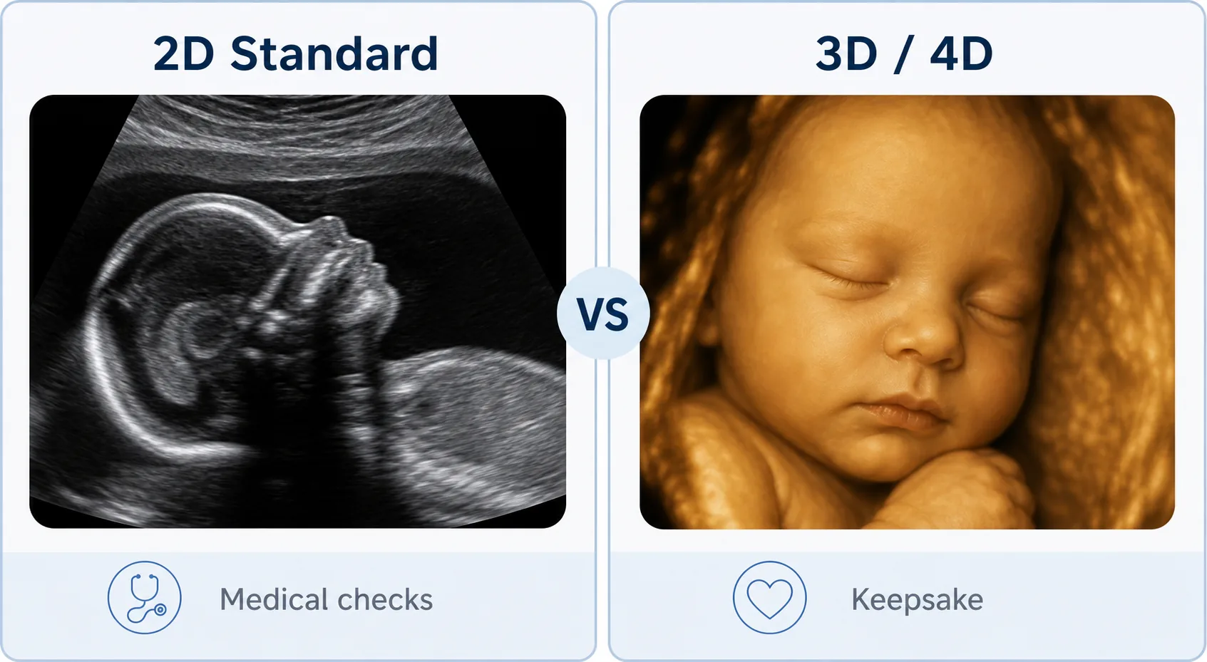
The medical anatomy scan uses 2D imaging, which gives the clearest view of internal structures for diagnosis. A 20-week 3D or 4D ultrasound produces the lifelike, keepsake-style pictures many parents recognise, but it is elective imaging, not a diagnostic assessment. The two serve different purposes, and the standard 2D scan is the one that checks your baby’s development.
What to Expect During a 20-Week Anatomy Scan?
The scan is painless and usually takes around 20 to 30 minutes, although it may take longer if your baby’s position makes certain views harder to capture. The sonographer may ask you to shift positions, roll slightly, or take a short break so they can complete the checklist.
Bladder instructions vary by provider, so follow the guidance in your appointment details. In some cases, you may be asked to arrive with a full or partially full bladder, while in others it may not be needed.
Bring your insurance card, appointment instructions, and any questions you want to ask about the scan or your baby’s development.
How Much Does a 20-Week Anatomy Scan Cost?
The cost of a 20-week anatomy scan depends on your location, insurance plan, provider, and whether the scan is part of routine prenatal care. Many patients have the anatomy scan covered as part of pregnancy care, but coverage and out-of-pocket costs can vary.
The safest step is to check with your clinic and insurance provider before the appointment. Ask whether the scan is considered routine prenatal care, whether it needs prior authorization, and what your expected cost may be.
If a repeat scan or specialist ultrasound is recommended, that may be billed differently from the routine anatomy scan, so it is worth confirming coverage before the follow-up visit.
FAQs
Related readingHow Many Prenatal Appointments Do You Have During Pregnancy?Read the guide →Frequently asked questions
What can be detected at a 20-week anatomy scan?
What are the 11 conditions checked at the 20-week scan?
What are good signs at 20 weeks pregnant?
Should I drink a lot of water before my 20-week anatomy scan?
What is the biggest indicator of Down syndrome?
Can the anatomy scan be done after 22 weeks?
Is the 20-week anatomy scan painful?
Is a 3D ultrasound the same as an anatomy scan?
Final Thoughts
The 20-week anatomy scan is an important pregnancy milestone because it gives your care team a detailed look at your baby’s development and helps guide the next steps in prenatal care.
Most results are reassuring, and when something needs a second look, your provider can walk you through what it means, what happens next, and which questions to ask before you leave.
You do not need to have every answer before the appointment. Bring your questions, ask what was checked, and let your care team help you understand the results in the context of your whole pregnancy.